
Hampton HR3-151 LCP Sandwich Set
LCP夹层集
应用
- Lipidic中间相膜蛋白的结晶筛选及二段法和分批法
特征
- 小体积LCP、双胶束和微间歇结晶
- 高质量玻璃光学
- 由于中间相团被物理地夹在两个光学清晰的表面之间,消除了中间相/水介质界面和相应的粗糙度,因此具有优良的滴光光学性能。
- 玻璃板允许使用交叉偏振片之间的无双折射检查来显示微晶。
- 超疏水玻璃表面
- 光场、紫外光和荧光显微镜的优化
- 稳定性好,耐蒸发
描述
LCP夹芯片组由一个底座玻璃滑块和一个优化的盖板滑块组成。LCP三明治集与著名的斯克里普斯研究所共同开发,位于美国加利福尼亚州拉霍拉,由保罗马里恩菲尔德(0890003)制造。
LCP玻璃夹芯片组是一种专门设计的平板,可用于96 Lipidic立方相矩阵筛选实验的自动或手动设置。本发明还可用于二段法和分批法。薄片(<2mm高)具有优良的光学性能,非常适合于微晶体的探测和交叉偏振片之间的无双折射成像。该平板允许在中间结晶试验中,50纳米色素,蛋白质/脂质中间相和1微升沉淀剂溶液每次试验。
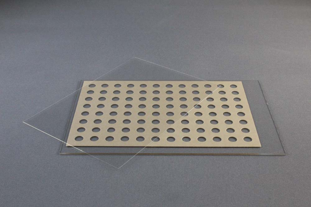
96井LCP玻璃夹层板由一个127.8×85.5x1mm玻璃基板和一个带有SBS兼容板足迹的140米厚的双粘性间隔板和一个0.2毫米厚的玻璃盖板组成。双粘垫片已粘在基板上。牛皮纸衬垫覆盖和保护双粘性间隔和基板顶部。0.2毫米厚的玻璃盖板适合和密封整个96井下板。另外,一系列的24硅化18x18mm正方形盖板(HR3-152)可以用来密封整个96井下板。每个18×18毫米1盖滑动密封4井。条形码的空间可用在板的左端。每口井可含立方相纳米粒子50粒,结晶试剂1微升。
18x18mm 1号硅化盖板由第一级水解级的抗化学腐蚀硼硅酸盐玻璃D 263 M制成。该幻灯片无色,清晰,适用于荧光显微镜。幻灯片的两侧都有一个超疏水的表面。幻灯片是没有的。厚度(0.13ro 0.16 mm)。幻灯片以两个隔间塑料箱提供,每箱100张幻灯片,每箱10盒,HR3-152型1,000张幻灯片。
碳化钨玻璃切割机可用于切割和拆卸密封LCP夹层套的上盖板。单独出售。
APPLICATIONS
- Crystallization screening of membrane proteins in lipidic mesophase as well as bicelle method and batch method
FEATURES
- Small volume LCP, bicelle and microbatch crystallization
- High quality glass optics
- Excellent drop optics since mesophase bolus is physically sandwiched between two optically clear surfaces eliminating the mesophase / aqueous medium interface and the corresponding roughness
- Glass plate allows for visualization of microcrystals using birefringence-free examination between crossed polarizers
- Superhydrophobic glass surfaces
- Optimized for bright field, UV and fluorescent microscopy
- Excellent stability, resistant to evaporation
DESCRIPTION
The LCP Sandwich Set consists of a base glass slide and an optimized cover slip. LCP Sandwich Set developed jointly with the renowned Scripps Research Institute in La Jolla, California, USA and is manufactured by Paul Marienfeld (0890003).
The LCP Glass Sandwich Set is a specially designed plate for either automated or manual setting of 96 Lipidic Cubic Phase matrix screening experiments. The plate can also be used for the bicelle method and batch method. The thin (< 2 mm high) plates have exquisite optical properties and are well suited for the detection of microcrystals and for birefringence-free imaging between crossed polarizers. The plates allow for in meso crystallization trials with 50 nanoliters protein/lipid mesophase and 1 microliter precipitant solution per trial.
The 96 well LCP Glass Sandwich Plate consists of a) 127.8 x 85.5 x 1 mm glass base plate with the footprint of an SBS-compliant plate, a 140 µm thick double sticky spacer with 96 punched out holes and b) a 0.2 mm thick glass coverslip. The double sticky spacer is already adhered to the base plate. A brown paper liner covers and protects the top of the double sticky spacer and base plate. The 0.2 mm thick glass coverslip fits over and seals the entire 96 well lower plate. Alternatively a series of twenty-four siliconized 18 x 18 mm No. 1 square cover slides (HR3-152) can be used to seal the entire 96 well lower plate. Each 18 x 18 mm No. 1 cover slide seals 4 wells. Space for a bar code is available at the left end of the plate. Each well can contain 50 nanoliters of cubic phase and 1 microliter of crystallization reagent.
18 x 18 mm No. 1 Siliconized Cover Slides are made from chemically resistant borosilicate glass D 263 M of the first hydrolytic class. The slides are colorless, clear, and suitable for fluorescence microscopy. The slides feature a super hydrophobic surface on both sides. The slides are No. thickness (0.13 ro 0.16 mm). The slides are supplied in a two compartment plastic box, 100 slides per box, 10 boxes per case, 1,000 slides total for HR3-152.
The Tungsten-carbide glass cutter can be used for cutting and removal of the upper coverglass that seals the LCP Sandwich Set. Sold separately.
| HR3-151 | LCP Sandwich Set | Pack of 20 |
| HR3-152 | 18 x 18 mm No. 1 Siliconized Cover Slides, Squares | Case of 10 packs、 |
ELATED ITEM(S)
REFERENCES
1. Nano-volume plates with excellent optical properties for fast, inexpensive crystallization screening of membrane proteins. Vadim Cherezov and Martin Caffrey. J. Appl. Cryst. (2003). 36, 1372-1377.
2. A robotic system for crystallizing membrane and soluble proteins in lipidic mesophases. Vadim Cherezov, Avinash Peddi, Lalitha Muthusubramaniam, Yuan F. Zheng and Martin Caffrey. Acta Cryst. (2004). D60, 1795-1807 DOI: 10.1107/S0907444904019109.
3. Bicelle crystallization: a new method for crystallizing membrane proteins yields a monomeric bacteriorhodopsin structure. Faham, S. & Bowie, J. U. (2002). J. Mol. Biol. 316, 1-6.