Hampton 带结晶loop环的底座(SPINE型) CrystalCap™ SPINE HT

Hampton 带结晶loop环的底座(SPINE型) CrystalCap™ SPINE HT

Hampton蛋白结晶试剂耗材
Hampton Research代理
欢迎访问Hampton Research官网或者咨询我们获取更多有关蛋白结晶试剂耗材产品信息。

Applications

Cryocrystallography   低温结晶学
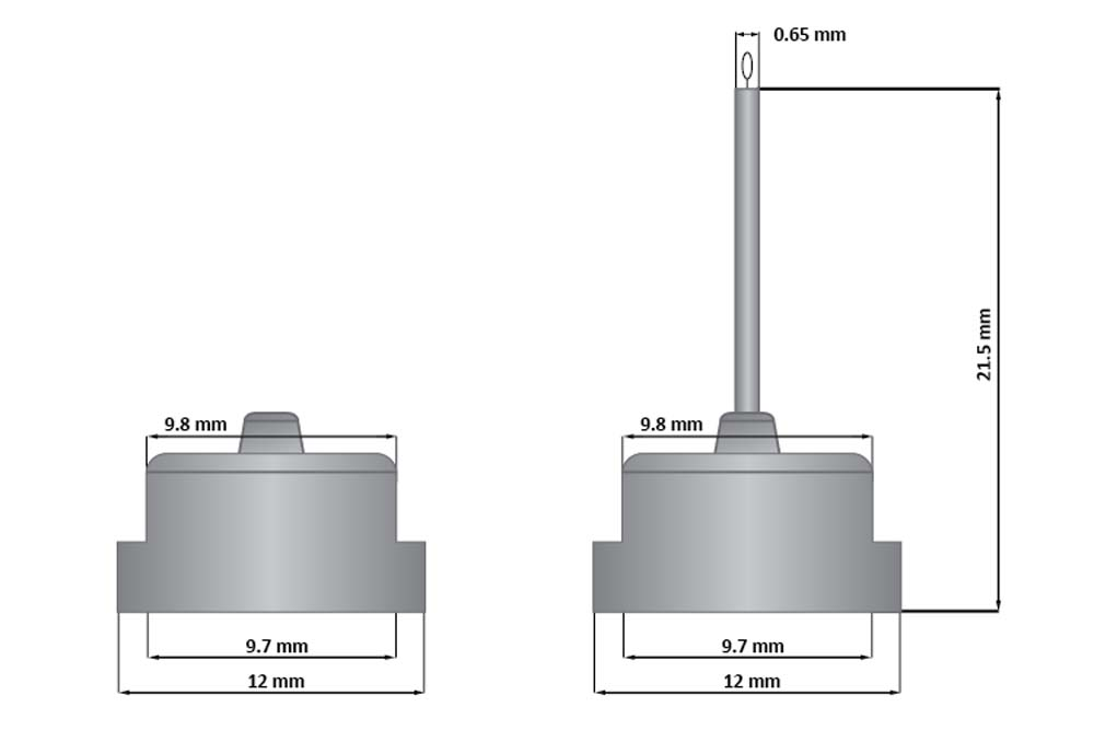
Features

SPINE style sample mount for cryocrystallography
CrystalCap SPINE HT attaches magnetically to CrystalCap Vial and Magnetic Goniometer Base     CrystalCap SPINE HT磁性地附着到CrystalCap小瓶和磁测角仪底座
Hollowed out design compatible with SPINE style grippers, auto mounters and sample handlers
2D alphanumeric coding for sample tracking & management
Bar coded, color coded, alphanumeric, magnetic cap
Color coded CryoLoop size
Description
The CrystalCap SPINE HT is a magnetic sample mount (also known as a cap, pin or goniometer base) designed for cryocrystallography systems that accept SPINE style caps. The CrystalCap SPINE HT attaches to a magnetic CrystalCap SPINE Vial and magnetic goniometer base. The tip of the CrystalCap SPINE HT accepts a Mounted CryoLoop™ or MicroTube™ fitted with a CryoLoop™.The CrystalCap SPINE Vial is a 1.8 ml cryo vial featuring two small vents and is compatible with the CrystalCap SPINE HT. A ring magnet is molded into the open end of the vial so that when the cap is positioned in the vial, the ring magnet holds the cap on the vial during cryogenic storage. The CrystalCap SPINE Vial features chamfered edges for enhanced cap positioning as well as a magnetic alloy bottom for stability. The HR8-114 CrystalCap SPINE Vial does have a magnet on the bottom of the vial.The hollowed out design of the CrystalCap SPINE HT is compatible with SPINE style grippers, auto mounters and sample handlers.The CrystalCap SPINE HT features a two dimensional (2D) alphanumeric 16 x 16 data matrix code on the underside of the cap. Each cap is also color coded for CryoLoop size.CrystalCap SPINE without 2D code/printing (HR8-094) is the CrystalCap SPINE HT cap only, without 2D code, without color code, without white white background on bottom of cap and no alphanumeric side labeling; cap only, without Mounted CryoLoop.Note: Caps with Mounted CryoLoops are sold without vials. Vials available separately.Color Coded Cap…………CryoLoop Size
Red…………………………….0.025-0.05 mm
Green…………………………….0.05-0.1 mm
Yellow……………………………..0.1-0.2 mm
Blue………………………………..0.2-0.3 mm
Blue/Red………………………….0.3-0.4 mm
Green/Red………………………..0.4-0.5 mm
Yellow/Red………………………..0.5-0.7 mm
Yellow/Green……………………..0.7-1.0 mmCompatible with the following Synchrotron Radiation BeamlinesNorth & South America
• The Advanced Light Source, Berkeley, California ALS 4.2.2, ALS 11.3.1, ALS 12.2.2, ALS 12.3.2
• The Advanced Photon Source, Argonne, Illinois APS 14-BM-C BioCARS, APS 14-ID-B BioCARS, APS 21-ID-D LS-CAT, APS 21-ID-E LS-CAT, APS 21-ID-F LS-CAT, APS 21-ID-G LS-CAT, APS 23-ID-B GA/CA, APS 23-ID-D GA/CA, APS 24-ID-E NE-CAT, APS 31-ID LR-CAT
• Center for Advanced Microstructures and Devices, Baton Rouge, Louisiana CAMD GCPCC
• Cornell High Energy, Synchrotron Source, Ithaca, New York CHESS A1, CHESS F1
• The Brazilian Synchrotron Light Laboratory, Sao Paulo, Brazil LNLS D03B-MX1, LNLS W01B-MX2
• National Synchrotron Light Source, Upton, New York NSLS X3B, NSLS X4A, NSLS X4C, NSLS X6A, NSLS X12B, NSLS X12C, NSLS X25, NSLS X26C, NSLS X29

Europe
• Cerdanyola del Vallés, Spain ALBA XALOC
• Berlin Electron Storage Ring Company for Synchrotron Radiation, Berlin, Germany BESSY 14.1, BESSY 14.2, BESSY 14.3
• Diamond Light Source, Didcot, Oxfordshire, England DIAMOND I02, DIAMOND I03, DIAMOND I04, DIAMOND I04.1, DIAMOND I24
• Elettra Sincrotrone Trieste, Trieste, Italy ELETTRA 5.2R
• European Molecular Biology Laboratory, Hamburg, Germany EMBL/DESY P13, EMBL/DESY P14
• European Synchrotron Radiation Facility, Grenoble, France ESRF BM14, ESRF BM30A, ID30A-1/MASSIF-1,
ID30A-3/MASSIF-3, ESRF ID23-1, ESRF ID23-2, ESRF ID29
• Max-Lab, Lund University, Sweden MAX II I911-2, MAX II I911-3
• Swiss Light Source at Paul Scherer Institut, Switzerland SLS X06DA-PXIII, SLS X06SA-PXI, SLS X10SA-PXII
• SOLEIL, Saint-Aubin, France SOLEIL PROXIMA1, SOLEIL PROXIMA2

Asia & Australia
• Shanghai Synchrotron Radiation Facility, Shanghai, China SSRF BL17U1
• Beijing Synchrotron Radiation Facility, Beijing, China BSRF 1W2B, BSRF 3W1A
• Super Photon ring-8 GeV, Japan SPRING-8 BL12B2, SPRING-8 BL24XU, SPRING-8 BL26B1, SPRING-8 BL26B2, SPRING-8 BL32B2, SPRING-8 BL32XU, SPRING-8 BL38B1, SPRING-8 BL41XU, SPRING-8 BL44B2, SPRING-8 BL44XU

CrystalCap SPINE HT

CrystalCap SPINE HT

CrystalCap SPINE HT

应用

  • 冰晶术

特征

  • 冰晶学用脊柱式样品架
  • 结晶帽脊骨HT磁性附着在晶体帽冰盖和磁干涉仪基座上
  • 与脊柱式夹持器、自动装配机和样本处理程序兼容的空心设计
  • 与自动示例处理程序兼容
  • 用于样本跟踪和管理的二维字母数字编码
  • 条形码,颜色编码,字母数字,磁帽
  • 彩色编码冷冻环尺寸

描述

结晶帽脊柱HT是一个磁性样品安装(也称为盖子,引脚或测角器底座),为接受脊柱式盖子的冰晶系统设计。该水晶帽脊柱HT附着在一个磁性水晶帽脊柱Vial和磁测角仪基座。水晶帽脊柱的尖端接受一个安装的冷冻环™。

水晶帽脊柱Vial是一个1.8毫升的冰瓶,有两个小通风口,并与水晶帽脊柱HT兼容。将环形磁铁模压到瓶的开口端,以便当盖被放置在瓶中时,环磁铁在低温储存期间将盖保持在瓶上。水晶帽脊柱的特点是倒角边缘,以加强帽子的定位,以及磁性合金底部的稳定性。HR8-114水晶帽脊椎骨在瓶底确实有一个磁铁。

水晶帽脊柱HT的空心设计与脊柱风格的夹持器、自动装配机和样品处理程序兼容。

水晶帽脊柱HT的特点是一个二维字母数字16×16数据矩阵代码在帽子的底部。每个帽子的颜色也编码为Cryoloop大小。

没有2D编码/打印的水晶帽脊柱(HR8-094)是水晶帽脊柱HT帽,没有2D代码,没有颜色代码,帽子底部没有白色背景,没有字母数字边标记;

注:装有深水瓶的瓶盖是不带瓶的。另外还有小瓶。

颜色编码,第.章,冷冻环尺寸
Red..0.025-0.05 mm
Green..0.05-0.1 mm
Yellow..0.1-0.2 mm
Blue..0.2-0.3 mm
Blue/Red..0.3-0.4 mm
绿/红.0.4-0.5毫米
Yellow/Red..0.5-0.7 mm
黄/绿.0.7至1.0毫米

与下列同步辐射光束线兼容

北美和南美洲
·高级光源,加州伯克利,ALS 4.2.2,ALS 11.3.1,ALS 12.2.2,ALS 12.3.2
*高级光子源,Argonne,伊利诺斯州APS 14-BM-C BioCARS,APS 14-ID-B BioCARS,APS 21-ID-D LS-CAT,APS 21-ID-E LS-CAT,APS 21-ID-F LS-CAT,APS 21-ID-G LS-CAT,APS 23-ID-B GA/CA,APS 23-ID-D GA/CA,APS 24 ID-E NE-CAT,APS 31 ID LR-CAT
·先进微结构和器件中心,巴吞鲁日,路易斯安那州CAMD GCPCC
·康奈尔高能,同步加速器源,伊萨卡,纽约国际象棋A1,国际象棋F1
*巴西同步光实验室,巴西圣保罗,LNLS D03B-MX1,LNLS W01B-MX2
国家同步光源II,厄普顿,纽约NSLS-II 17-ID-1 AMX和NSLS-II 17-ID-2 FMX。NSLs-II 19-ID NYX

欧洲
·Cerdanyola del Vallés,西班牙Alba XALOC
·柏林同步辐射电子储存环公司,德国柏林BESSY 14.1、BESSY 14.2、BESSY 14.3
·钻石光源,迪科特,牛津郡,英国钻石I02,钻石I 03,钻石I 04,钻石I 04.1,钻石I24
·Elettra Sincrotrone里雅斯特,意大利里雅斯特
·欧洲分子生物学实验室,德国汉堡EMBL/DESY P13、EMBL/DESY P14
欧洲同步辐射设施,法国格勒诺布尔,法国ESRF BM 14,ESRF BM30A,ID30A-1/Massif-1,ID30A-3/Massif-3,ESRF ID23-1,ESRF ID23-2,ESRF ID29
·瑞典隆德大学实验室,Max II I 911-2,Max II I 911-3
·瑞士Paul Scherer研究所的瑞士光源,瑞士SLS X06DA-PXIII,SLS X06SA-PXI,SLS X10SA-PXII
太阳、圣奥宾、法国太阳PROXIMA 1、太阳PROXIMA 2

亚洲和澳大利亚
·上海同步辐射装置,中国上海SSRF BL17U1
·北京同步辐射设施,北京,中国北京BSRF 1W2B,BSRF 3W1A
超级光子环-8 GeV,日本Spring-8 BL12B2,Spring-8 BL24XU,Spring-8 BL26B1,Spring-8 BL26B2,Spring-8 BL32B2,Spring-8 BL32XU,Spring-8 BL38B1,Spring-8 BL41XU,Spring-8 BL44B2,Spring-8 BL44XU

CAT NO NAME DESCRIPTION
HR8-118 CrystalCap SPINE HT 0.025-0.05 mm CryoLoop, without Vial – 30 pack
HR8-120 CrystalCap SPINE HT 0.05-0.1 mm CryoLoop, without Vial – 30 pack
HR8-122 CrystalCap SPINE HT 0.1-0.2 mm CryoLoop, without Vial – 30 pack
HR8-124 CrystalCap SPINE HT 0.2-0.3 mm CryoLoop, without Vial – 30 pack
HR8-126 CrystalCap SPINE HT 0.3-0.4 mm CryoLoop, without Vial – 30 pack
HR8-128 CrystalCap SPINE HT 0.4-0.5 mm CryoLoop, without Vial – 30 pack
HR8-130 CrystalCap SPINE HT 0.5-0.7mm CryoLoop, without Vial – 30 pack
HR8-132 CrystalCap SPINE HT 0.7-1.0 mm CryoLoop, without Vial – 30 pack
HR8-112 CrystalCap SPINE HT Cap only – 30 pack
HR8-116 CrystalCap SPINE HT Cap/Vial Cap/Vial only – 30 pack
HR8-094 CrystalCap SPINE without 2D code/printing Cap only – 30 pack
HR8-114 CrystalCap SPINE Vial Vial only – 30 pack

RELATED ITEM(S)

Hampton HR2-591 PEG 3350 聚乙二醇3350

Hampton HR2-591 PEG 3350 聚乙二醇3350

Hampton HR2-591 PEG 3350 Polyethylene glycol 3,350 Monodisperse

Hampton research蛋白结晶试剂代理–上海金畔生物
代理优势:货期3-4周。
欢迎访问Hampton research官网或者咨询我们获取更多产品信息。

Optimize Reagents > Optimize – Polymers > Polyethylene glycol 3,350 Monodisperse

APPLICATIONS

  • Crystallization Polyethylene glycol 3,350 for formulating screens or for optimization

聚乙二醇3,350单分散

应用

  • 结晶聚乙二醇3,350用于筛选或优化

FEATURES

  •  Sterile filtered solution
  • Formulated in Type 1+ ultrapure water: 18.2 megaohm-cm resistivity at 25°C, < 5 ppb Total Organic Carbon, bacteria free (<1 Bacteria (CFU/ml)), pyrogen free (<0.03 Endotoxin (EU/ml)), RNase-free (< 0.01 ng/mL) and DNase-free (< 4 pg/µL)

特征

  • 无菌过滤液
  • 在1+超纯水中配制:在25°C下电阻率为18.2兆,总有机碳<5 ppb,无细菌(<1细菌(CFU/ml)),无热原(<0.03内毒素(EU/ml)),无RNase(<0.01 ng/mL)和无DNase(<4 pg/L)

DESCRIPTION   描述

United States Pharmacopeia (USP)/National Formulary (NF), Pharmacopoea Europaea (Ph. Eur.), and Food Chemicals Codex (FCC) grade PEG 3350. For research use only. Not for diagnostic use.

Polyethylene glycol 3,350
Synonyms: PEG 3,350
H(OCH2CH2)nOH
Average Mr 3015-3685
CAS Number [25322-68-3]
EC Number 500-038-2
Merck 14,7568
MDL Number MFCD00081839
PubChem Substance ID 24898432
Polydispersity Index 1.05-1.10 (Polydispersity is a measure of the molecular weight distribution of a polymer. It is equal to Mw/Mn. That is to say, “weight-average molecular weight” divided by “number average molecular weight”.)

Aldehyde: None detected
Peroxide: None detected

美国药典(USP)/国家配方集(NF)、欧洲药典(Ph.EUR.)和食品化学品法典(FCC)级PEG 3350。只供研究用。不是用来诊断的。

聚乙二醇3,350
同义词:PEG 3,350
H(Och)22)n
平均Mr 3015-3685
核证机关编号[25322-68-3]
教统会编号500-038-2
默克14,7568
MDL号码MFCD 00081839
PubChem物质ID 24898432
多分散性指数1.05~1.10(多分散性是衡量聚合物分子量分布的指标。它等于Mw/Mn。也就是说,“重量-平均分子量”除以“数平均分子量”)。

醛:没有检测到
过氧化物:没有检测到

HR2-527测量电导率范围:44.0-55.1 S/cm,25°C
HR2-527测量折射率范围:1.40137~1.40213℃,20°C

HR2-527 Measured Conductivity Range: 44.0 – 55.1 µS/cm at 25°C
HR2-527 Measured Refractive Index Range: 1.40137 – 1.40213 at 20°C

HR2-591 Polyethylene glycol 3,350 Monodisperse 500 grams
HR2-527 50% w/v Polyethylene glycol 3,350 Monodisperse 200 mL

REFERENCES  参考文献

1. Crystal structure of a Soluble form of the human T Cell coreceptor CD8 at 2.6 A resolution. Leahy, DJ; Axel, R; Hendrickson, WA. Cell 68, 1145- 1162, 1992.

2. McPherson, A. 1976, Crystallization of proteins from polyethylene glcyol. J. Biol. Chem. 251: 6300.

Hampton HR4-729 CrystalWand™ Magnetic 磁力结晶杆

Hampton HR4-729 CrystalWand™ Magnetic磁力结晶杆

Applications

CrystalCap Magnetic & CrystalCap Copper Magnetic handling tool

Description
The CrystalWand Magnetic is designed to be used exclusively with the CrystalCap Magnetic, CrystalCap Copper Magnetic, and CrystalCap HT systems during transfer of the caps from the vial to the goniometer head and from the goniometer to the vial. The 6 1/2” (165 mm) long chrome plated steel wand features a plastic housing enclosing a spring tensioned plunger that when depressed, moves a non-magnetic steel platform away from the magnet housed in the end of the wand. This causes the steel CrystalCap to detach from the wand and be placed readily into the vial. When the platform is retracted, the wand securely holds the steel CrystalCap Magnetic.

描述

水晶棒磁性是专门用于水晶帽,水晶帽ALS,和水晶帽脊柱系统期间,帽子从小瓶转到测角仪头和从测角仪到小瓶。该6 1/2“(165毫米)长镀铬钢棒的特点是一个塑料外壳包围弹簧拉力柱塞,当下降时,移动一个非磁性钢平台,远离磁铁住在魔杖的末端。这导致钢水晶帽从魔杖上分离出来,很容易被放入瓶子中。当平台被收回时,魔杖牢固地握住钢水晶帽磁性。

CAT NO NAME DESCRIPTION
HR4-729 CrystalWand Magnetic, Straight each

RELATED ITEM(S)
CrystalCap™
CrystalCap™ SPINE HT
CrystalCap™ ALS HT
CrystalCap™ ALS

图片仅供参考,请以实物为准。
正文中列出的所有试剂只能用于测试或研究,不能作为”药品”,”食品”,”家庭用品”等使用。
我司所销售的化学试剂、原料等所有产品(包括但不限于抗生素类、蛋白质类、试剂盒类产品等)仅限用于科学研究用途,不得作用于人体。

Hampton HR4-710 Izit Crystal Dye 蛋白染色试剂

Hampton HR4-710 Izit Crystal Dye 蛋白染色试剂

Hampton research蛋白结晶试剂代理–上海金畔生物

代理优势:货期4周左右。

欢迎访问Hampton research官网或者咨询我们获取更多产品信息。

Applications
Differentiate protein crystals from salt crystals
Features
0.22 micron sterile filtered solution
Small molecule dye penetrates solvent channels of macromolecular crystals, coloring the crystals blue
Salt crystals cannot absorb Izit and remain colorless
Differentiate protein and non-protein based phase separation
Formulated in Type 1+ ultrapure water: 18.2 megaohm-cm resistivity at 25掳C, < 5 ppb Total Organic Carbon, bacteria free (<1 Bacteria (CFU/ml)), pyrogen free (<0.03 Endotoxin (EU/ml)), RNase-free (< 0.01 ng/mL) and DNase-free (< 4 pg/碌L)

Izit coloring protein phase separation blue

Crystals before Izit

Crystals after Izit

Description
A crystal! Is it protein or is it salt? Have you ever asked yourself this question? Sure you could mount the crystal in question and take a quick look at the diffraction pattern but perhaps you’re too lazy or you don’t have an x-ray facility. Well, there’s always the crush test. One simply takes a MicroTool™ or Crystal Probe™ and crushes the suspect crystal. A click or solid crunch is indicative of salt while a powder or silent destruction might indicate one just destroyed a perfectly good protein crystal. Izit is here to help. Simply place 1 µl of Izit in the sample drop and wait for an hour or so. Izit is a small molecule dye which will fill the solvent channels in protein crystals, coloring the crystals blue. With the appropriate dilution, Izit will in fact leave a clear drop with blue crystals, as illustrated in the picture above. Salt crystals do not possess these large solvent channels. Therefore, Izit cannot enter the crystal, leaving one with a clear crystal and a blue drop. Izit is especially nice for small microcrystals or questionable precipitate. The 0.5 ml vial of Izit is sufficient for more than thousands of crystallization drops.

Each Izit vial contains 0.5 ml of Izit dye. All solutions are formulated using ultra-pure water and are sterile filtered. Crystallization accessories are sold separately.

应用
区分蛋白质晶体和盐晶体
特征
0.22微米无菌过滤溶液
小分子染料渗透大分子晶体的溶剂通道,使晶体呈蓝色
盐晶体不能吸收 Izit 并保持无色
区分基于蛋白质和非蛋白质的相分离
在 1+ 型超纯水中配制:25°C 时电阻率为 18.2 兆欧-厘米,< 5 ppb 总有机碳,无细菌(<1 细菌 (CFU/ml)),无热原(<0.03 内毒素 (EU/ml)) , 无 RNase (< 0.01 ng/mL) 和无 DNase (< 4 pg/mL)
Izit 着色蛋白质相分离蓝色

伊兹特之前的水晶

伊兹特之后的水晶

描述
一个水晶!是蛋白质还是盐?你有没有问过自己这个问题?当然,您可以安装有问题的晶体并快速查看衍射图,但也许您太懒了,或者您没有 X 射线设备。好吧,总是有粉碎测试。只需使用 MicroTool™ 或 Crystal Probe™ 即可粉碎可疑晶体。咔嗒声或固体嘎吱声表示盐分,而粉末或无声破坏可能表示刚刚破坏了完美的蛋白质晶体。 Izit 是来帮忙的。只需将 1 µl Izit 放入样品滴中并等待一个小时左右。 Izit 是一种小分子染料,可填充蛋白质晶体中的溶剂通道,使晶体呈蓝色。通过适当的稀释,Izit 实际上会留下带有蓝色晶体的清晰液滴,如上图所示。盐晶体不具有这些大的溶剂通道。因此,伊兹特无法进入水晶,留下一个透明的水晶和蓝色的水滴。 Izit 特别适用于小微晶或可疑沉淀物。 0.5 毫升的 Izit 小瓶足以容纳超过数千滴结晶滴。

每个 Izit 小瓶含有 0.5 毫升 Izit 染料。所有溶液均使用超纯水配制并经过无菌过滤。结晶配件单独出售。

CAT NO NAME DESCRIPTION
HR4-710 Izit Crystal Dye 0.5 ml vial

Hampton HR4-670夹钳-直 Vial Clamp – Straight

Hampton HR4-670夹钳-直 Vial Clamp – Straight

Applications
Vial support and manipulation
Features
Straight end tip
Hemostat style closure
Description
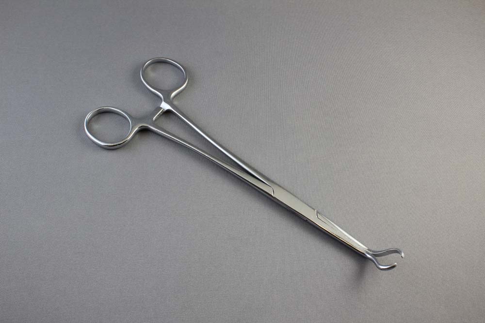
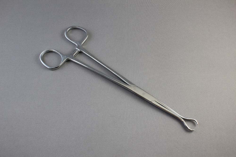
The Vial Clamp™ – Straight is a chrome plated, hemostat style tool. It has a tip shaped to hold the storage vial of all the CrystalCap Systems™ straight when the clamp is closed. The clamp can be locked using the hemostat style closure. This clamp makes it easy to dip the storage vial into liquid nitrogen for crystal storage. The complete length of the clamp is straight, with an overall length of 195 mm. When the vial is placed in the clamp, the length of it is positioned perpendicular, 90° to the length of the clamp.

Vial Clamp™ – Curved
Images
Vial Clamp™ – Straight
Images

Hampton HR4-670夹钳-直 Vial Clamp – Straight

CAT NO NAME DESCRIPTION
HR4-670 Vial Clamp  Straight each

Hampton CrystalCap的磁力支架 Magnetic Base

Hampton CrystalCap的磁力支架 Magnetic Base

FEATURES

  • Available in 2 strengths
  • Magnetic support for CrystalCap

Hampton蛋白结晶试剂耗材
Hampton Research代理
欢迎访问Hampton Research官网或者咨询我们获取更多有关蛋白结晶试剂耗材产品信息。


DESCRIPTION

The Magnetic Base (9.3 mm diameter support) is a machined brass piece fitted with a magnet. The base fits into the goniometer head, providing a secure base for the CrystalCap System™ sample holders. Fits most goniometer heads. The Magnetic Base is available in two strengths: strong and medium. The stem of the magnet is 3 mm in diameter. The length of the stem is 5 mm. The thickness of the platform is 3.6 mm.

特征

  • 有两种优势
  • 晶帽磁性支架

描述

磁性基座(直径9.3毫米)是一个装有磁铁的加工黄铜件。该底座适合于测角仪头,为水晶帽系统™样品保持架提供了一个安全的底座。适合大多数测角头。磁基有两种优势:强的和中等的。磁铁的阀杆直径为3毫米。茎长5mm。平台厚度为3.6mm。

HR4-943 Magnetic Base – Strong 9,000 gauss – each
HR4-627 Magnetic Base – Medium 6,000 gauss – each

RELATED ITEM(S)

Hampton 盖玻片切割刀

Hampton 盖玻片切割刀

Hampton Tungsten Carbide Scribe & Glass Cutter with Replaceable Tips

碳化钨刀头&可更换刀尖的玻璃切割机

APPLICATIONS  应用

  • Useful for scribing, cutting and removal of the upper coverglass that seals the LCP Sandwich Set.
  • 适用于划线,切割和拆卸上盖板,密封LCP三明治套。

FEATURES  特征

  • Machined, lightweight aluminum handle
  • Replaceable tungsten carbide scribe tip
  • Diamond knurling for comfortable and sure grip
  • Pocket clip
  • Size: 8 mm diameter, 130 mm length
  • Reversible tip protects tip and user
  • 机加工的,轻铝手柄
  • 可更换碳化钨针尖
  • 钻石滚花舒适可靠的抓地力
  • 袖珍夹
  • 尺寸:直径8毫米,长度130毫米
  • 可逆提示保护提示和用户

DESCRIPTION

Tungsten Carbide scribe and glass cutter with replaceable tungsten carbide tip. Tungsten Carbide Tip can be removed and inverted to protect tip and user. Knurled aluminum alloy body and pocket clip. Overall length 130 mm. Useful for cutting and removal of the upper coverglass that seals the HR3-151 LCP Sandwich Set.

Tungsten carbide scribe tips are not threaded and are held in place with a threaded tip chuck. To remove tip, turn the tip chuck counterclockwise one turn (without completely removing the chuck) while holding the aluminum handle stationary, and pull the tip from the chuck and handle. To install a new tip, slide the new tip into the chuck and handle and tighten the tip chuck clockwise to secure the new tip into the handle. Loosen tip chuck, remove tip, reserve and install with the tip inverted to protect user and the tip when not in use.

Tungsten Carbide Scribe is supplied with one tip installed. Replacement tips sold separately. Tungsten Carbide Scribe Tip is a set of 3 replacement tips designed for the Tungsten Carbide Scribe HR4-702. Tungsten carbide Tip in steel shank to strengthen and protect carbide from damage. Scribe Tip Diameter x Length: 3 mm X 32 mm.

See the JOVE video for more details about using the Tungsten Carbide glass cutter to open LCP Sandwich Set experiments:

描述

碳化钨切割机和可更换碳化钨针尖的玻璃切割机。碳化钨针尖可以拆卸和倒置,以保护针尖和使用者。滚花铝合金阀体和口袋夹。总长130毫米。用于切割和移除上盖板,密封HR3-151 LCP夹层套。

碳化钨刀尖不是螺纹的,而是用螺纹夹头固定在适当的位置。若要移除夹头,请在保持铝制手柄固定的同时逆时针旋转一次(不完全移除卡盘),并从卡盘和手柄上拉出夹头。若要安装新的尖端,请将新的尖端滑动到卡盘中,并按顺时针方向拧紧夹头卡盘,以将新的尖端固定到手柄中。松开刀尖卡盘,拆下针尖,保留并安装倒装,以保护使用者和未使用时的针尖。

碳化钨Scribe是提供与一个尖端安装。更换提示单独出售。碳化钨刀尖是为碳化钨Scribe HR4-702设计的一套3种替代尖端。钢柄碳化钨针尖强化和防止碳化物损坏。划线头直径x长度:3毫米x 32毫米。

HR4-702 Tungsten Carbide Scribe 笔套 Each
HR4-703 Tungsten Carbide Scribe Tip 笔头 Pack of 3 tips

RELATED ITEM(S)

REFERENCES

1. Li, D., Boland, C., Walsh, K., Caffrey, M. Use of a Robot for High-throughput Crystallization of Membrane Proteins in Lipidic Mesophases. J. Vis. Exp. (67), e4000, DOI: 10.3791/4000 (2012).

2. Cherezov, V., and M. Caffrey. (2003) Nano-volume plates with excellent optical properties for fast, inexpensive crystallization screening of membrane proteins. J. Appl. Cryst. 36: 1372-1377.

3. Cherezov, V., Peddi, A., Muthusubramaniam, L., Zheng, Y.F., and M. Caffrey. (2004) A robotic system for crystallizing membrane and soluble proteins in lipidic mesophases. Acta Crystallogr. D Biol. Crystallogr. 60: 1795-1807.

4. Caffrey, M., Li, D., & Dukkipati, A. Membrane protein structure determination using crystallography and lipidic mesophases – recent advances and successes. Biochemistry. DOI: 10.1021/bj300010w, 2012.

Hampton Capillary Cutting Stone

Hampton Capillary Cutting Stone

APPLICATIONS

  • The easiest way to cut capillaries

FEATURES

  • Score and snap.
  • Ceramic cutting stone

DESCRIPTION

These small (25 mm x 25 mm x 0.65 mm) cutting stones are used to make clean cuts of glass and quartz capillaries. Use the edge of the stone to etch the capillary then gently snap the capillary in two. This is useful for cutting the tips off closed capillaries used for mounting single crystals for x-ray diffraction analysis or for cutting capillaries to the desired length.

Capillary Cutting Procedure

1. Gently place the capillary on a clean, flat surface.
2. Gently holding the stone at approximately 30° angle to the capillary tube, starting near one edge of the stone, proceeding in a single, smooth motion, draw the length of the non-serrated edge of the stone across the capillary one time . Apply just enough pressure to score the capillary, do not attempt to cut through the capillary.
3. Pull the capillary axially until it breaks. If it won’t break, the score was not deep enough. Repeat the above steps, pressing down with slightly more force while drawing the cleaving stone the capillary.
4. Once cut, inspect the end finish to ensure the cut quality meets the application requirements.

毛细管切割石材™

应用

  • 切毛细血管最简单的方法

特征

  • 分分秒秒。
  • 陶瓷切割石

描述

这些小切割石(25毫米x25毫米x 0.65毫米)被用来切割玻璃和石英毛细血管。用石头的边缘蚀刻毛细,然后轻轻地把毛细拧成两半。这是有用的切尖关闭关闭毛细管用于安装单晶x射线衍射分析或切割毛细血管到所需的长度。

毛细管切割程序

1.轻轻地将毛细放在干净、平坦的表面上。
2.以大约30°的角度轻轻握住毛细管,从石的边缘开始,以一次平滑的方式进行,一次画出石的非锯齿状边缘的长度。施加足够的压力来打分毛细血管,不要试图切断毛细血管。
3.轴向拉毛,直至毛细血管断裂。如果它不能打破,比分是不够深的。重复上面的步骤,用稍微大一点的力量按压下来,同时把切割的石头拉到毛细上。
4.切割后,检查成品,确保切割质量符合使用要求。

RELATED IT

HR4-334 Capillary Cutting Stone each、

EM(S)

Hampton HR3-151 LCP Sandwich Set

Hampton HR3-151 LCP Sandwich Set

LCP夹层集

应用

  • Lipidic中间相膜蛋白的结晶筛选及二段法和分批法

特征

  • 小体积LCP、双胶束和微间歇结晶
  • 高质量玻璃光学
  • 由于中间相团被物理地夹在两个光学清晰的表面之间,消除了中间相/水介质界面和相应的粗糙度,因此具有优良的滴光光学性能。
  • 玻璃板允许使用交叉偏振片之间的无双折射检查来显示微晶。
  • 超疏水玻璃表面
  • 光场、紫外光和荧光显微镜的优化
  • 稳定性好,耐蒸发

描述

LCP夹芯片组由一个底座玻璃滑块和一个优化的盖板滑块组成。LCP三明治集与著名的斯克里普斯研究所共同开发,位于美国加利福尼亚州拉霍拉,由保罗马里恩菲尔德(0890003)制造。

LCP玻璃夹芯片组是一种专门设计的平板,可用于96 Lipidic立方相矩阵筛选实验的自动或手动设置。本发明还可用于二段法和分批法。薄片(<2mm高)具有优良的光学性能,非常适合于微晶体的探测和交叉偏振片之间的无双折射成像。该平板允许在中间结晶试验中,50纳米色素,蛋白质/脂质中间相和1微升沉淀剂溶液每次试验。

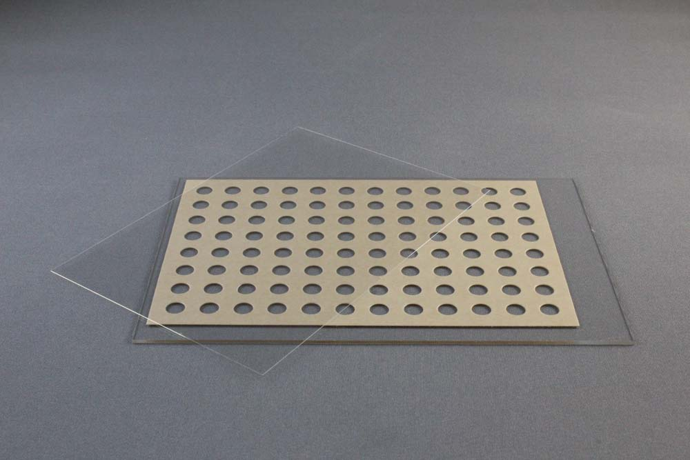
96井LCP玻璃夹层板由一个127.8×85.5x1mm玻璃基板和一个带有SBS兼容板足迹的140米厚的双粘性间隔板和一个0.2毫米厚的玻璃盖板组成。双粘垫片已粘在基板上。牛皮纸衬垫覆盖和保护双粘性间隔和基板顶部。0.2毫米厚的玻璃盖板适合和密封整个96井下板。另外,一系列的24硅化18x18mm正方形盖板(HR3-152)可以用来密封整个96井下板。每个18×18毫米1盖滑动密封4井。条形码的空间可用在板的左端。每口井可含立方相纳米粒子50粒,结晶试剂1微升。

18x18mm 1号硅化盖板由第一级水解级的抗化学腐蚀硼硅酸盐玻璃D 263 M制成。该幻灯片无色,清晰,适用于荧光显微镜。幻灯片的两侧都有一个超疏水的表面。幻灯片是没有的。厚度(0.13ro 0.16 mm)。幻灯片以两个隔间塑料箱提供,每箱100张幻灯片,每箱10盒,HR3-152型1,000张幻灯片。

碳化钨玻璃切割机可用于切割和拆卸密封LCP夹层套的上盖板。单独出售。

APPLICATIONS

  • Crystallization screening of membrane proteins in lipidic mesophase as well as bicelle method and batch method

FEATURES

  • Small volume LCP, bicelle and microbatch crystallization
  • High quality glass optics
  • Excellent drop optics since mesophase bolus is physically sandwiched between two optically clear surfaces eliminating the mesophase / aqueous medium interface and the corresponding roughness
  • Glass plate allows for visualization of microcrystals using birefringence-free examination between crossed polarizers
  • Superhydrophobic glass surfaces
  • Optimized for bright field, UV and fluorescent microscopy
  • Excellent stability, resistant to evaporation

DESCRIPTION

The LCP Sandwich Set consists of a base glass slide and an optimized cover slip. LCP Sandwich Set developed jointly with the renowned Scripps Research Institute in La Jolla, California, USA and is manufactured by Paul Marienfeld (0890003).

The LCP Glass Sandwich Set is a specially designed plate for either automated or manual setting of 96 Lipidic Cubic Phase matrix screening experiments. The plate can also be used for the bicelle method and batch method. The thin (< 2 mm high) plates have exquisite optical properties and are well suited for the detection of microcrystals and for birefringence-free imaging between crossed polarizers. The plates allow for in meso crystallization trials with 50 nanoliters protein/lipid mesophase and 1 microliter precipitant solution per trial.

The 96 well LCP Glass Sandwich Plate consists of a) 127.8 x 85.5 x 1 mm glass base plate with the footprint of an SBS-compliant plate, a 140 µm thick double sticky spacer with 96 punched out holes and b) a 0.2 mm thick glass coverslip. The double sticky spacer is already adhered to the base plate. A brown paper liner covers and protects the top of the double sticky spacer and base plate. The 0.2 mm thick glass coverslip fits over and seals the entire 96 well lower plate. Alternatively a series of twenty-four siliconized 18 x 18 mm No. 1 square cover slides (HR3-152) can be used to seal the entire 96 well lower plate. Each 18 x 18 mm No. 1 cover slide seals 4 wells. Space for a bar code is available at the left end of the plate. Each well can contain 50 nanoliters of cubic phase and 1 microliter of crystallization reagent.

18 x 18 mm No. 1 Siliconized Cover Slides are made from chemically resistant borosilicate glass D 263 M of the first hydrolytic class. The slides are colorless, clear, and suitable for fluorescence microscopy. The slides feature a super hydrophobic surface on both sides. The slides are No. thickness (0.13 ro 0.16 mm). The slides are supplied in a two compartment plastic box, 100 slides per box, 10 boxes per case, 1,000 slides total for HR3-152.

The Tungsten-carbide glass cutter can be used for cutting and removal of the upper coverglass that seals the LCP Sandwich Set. Sold separately.

HR3-151 LCP Sandwich Set Pack of 20
HR3-152 18 x 18 mm No. 1 Siliconized Cover Slides, Squares Case of 10 packs、

ELATED ITEM(S)

REFERENCES

1. Nano-volume plates with excellent optical properties for fast, inexpensive crystallization screening of membrane proteins. Vadim Cherezov and Martin Caffrey. J. Appl. Cryst. (2003). 36, 1372-1377.

2. A robotic system for crystallizing membrane and soluble proteins in lipidic mesophases. Vadim Cherezov, Avinash Peddi, Lalitha Muthusubramaniam, Yuan F. Zheng and Martin Caffrey. Acta Cryst. (2004). D60, 1795-1807 DOI: 10.1107/S0907444904019109.

3. Bicelle crystallization: a new method for crystallizing membrane proteins yields a monomeric bacteriorhodopsin structure. Faham, S. & Bowie, J. U. (2002). J. Mol. Biol. 316, 1-6.